
Ciliates Associated with Abalone
On this page
Category
Category 4 (Negligible Regulatory Significance in Canada)
Common, generally accepted names of the organism or disease agent
Ciliates of abalone
Scientific name or taxonomic affiliation
Mantoscyphidia sp., Scyphidia-like and Sphenophrya-like ciliates. Numerous species of ciliates in the orders Thigmotrichida, Peritrichida, Heterotrichida and Hypotrichida have been reported to live in close association with marine molluscs (Lauckner 1983).
Geographic distribution
Probably ubiquitous with reports of Mantoscyphidia sp. from abalone in southern Africa and similar looking ciliates in abalone in British Columbia, Canada and Baja California, Mexico.
Host species
Haliotis midae, Haliotis spadicea, Haliotis kamtschatkana, Haliotis rufescens, Haliotis iris and probably other species of abalone.
Impact on the host
Large numbers elicit no obvious host-response and attachment to the gills, mantle cavity or esophageal pouch epithelium appears to be superficial. Some populations of Mantoscyphidia sp. in abalone had attached ectosymbiotic ellobiophryid ciliates with no apparent adverse affects to the host ciliate or abalone (H. midae and H. spadicea) (Botes et al. 1998). Ciliates similar in morphology to Mantoscyphidia sp. were observed in the esophageal pouches of cultured juvenile H. kamtschatkana held in tanks for several months with an inadequate supply of food in British Columbia, Canada. Caceres-Martinez and Tinoco-Orta (2001) suggested that the ciliates in the esophageal pouches of cultured H. rufescens in Baja California may be commensal symbionts because prevalence and total numbers were similar in healthy and moribund abalone but indicated that further studies are required to determine the nature of the symbiosis.
Diagnostic techniques
Smears
Check smears of the gills under a microscope for free-swimming ciliates measuring 30-60 µm in length.
Histology
Ciliates lie on or in-between the gill lamellae, have a large densely basophilic macronucleus and a smaller posteriorly located micronucleus. The body is pyriform in shape with a pointed anterior end. Attachment is superficial and there is no evidence of tissue penetration.
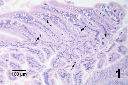
Figure 1. Numerous mantoscyphidian-like ciliates (arrows) in the esophageal pouches of cultured juvenile Haliotis kamtschatkana held in tanks for several months with an inadequate supply of food. Haematoxylin and eosin stain.
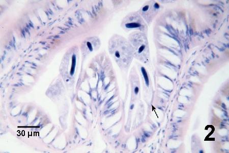
Figure 2. Several mantoscyphidian-like ciliates within a fold of the esophageal pouch of the same specimen as in Fig.1. There is no evidence of pathology to the abalone even at the attachment site of the ciliate (arrow). Haematoxylin and eosin stain.
Methods of control
Prevention and control impractical. These ciliates are ubiquitous.
References
Botes, H., L. Basson and J.G. Van As. 1998. Ectosymbiotic ellobiophryid peritrichs associated with abalone. Proceeding of the Microscopy Society of Southern Africa 28: 75.
Caceres-Martinez, J. and G.D. Tinoco-Orta. 2001. Symbionts of cultured red abalone, Haliotis rufescens from Baja California, Mexico. Journal of Shellfish Research 20: 875-881.
Diggles, B.K. and M. Oliver. 2005. Diseases of cultured paua (Haliotis iris) in New Zealand. In: Walker, P.J., R.G. Lester, M.G. Bondad-Reantaso (eds.) Diseases in Asian Aquaculture V. Proceedings of the 5th Symposium on Diseases in Asian Aquaculture. Fish Health Section, Asian Fisheries Society, Manila. pp. 275-287.
Lauckner, G. 1983. Diseases of Mollusca: Bivalvia. In: O. Kinne (ed.). Diseases of Marine Animals. Volume II: Introduction, Bivalvia to Scaphopoda. Biologische Anstalt Helgoland, Hamburg, p. 732-750.
Citation Information
Bower, S.M. (2006): Synopsis of Infectious Diseases and Parasites of Commercially Exploited Shellfish: Ciliates Associated with Abalone.
Date last revised: December 2006
Comments to Susan Bower
- Date modified: