
Ciliates Associated with Sea Urchins
On this page
Category
Category 4 (Negligible Regulatory Significance in Canada)
Common, generally accepted names of the organism or disease agent
Ciliates of sea urchins
Scientific name or taxonomic affiliation
Unidentified species of ciliates have been observed in close association with sea urchins tissues.
Geographic distribution
Probably ubiquitous but each species may have a limited geographic range.
Host species
Strongylocentrotus droebachiensis, Strongylocentrotus franciscanus and probably other species of sea urchins.
Impact on the host
No obvious host-response nor tissue damage despite the close association with the epithelium of the intestinal tract and external integument. Two morphological types of ciliates were frequently observed (prevalences of about 35% in some locations) embedded within epithelial folds in the digestive tract of sea urchins from the Pacific and Atlantic coasts of Canada. Dual infections with both types of ciliates have been observed. Trichodinids were common adjacent to the outer surface of the test of sea urchins from some locations (75% prevalence in S. droebachiensis from Malaspina Inlet, British Columbia, Canada and 80% prevalence in the same species of sea urchin from Canso, Nova Scotia, Canada).
Diagnostic techniques
Histology
Ciliates adjacent to the epithelial surface with no evidence of damage to host tissues. Ciliate Type A was elongate, spindle-shaped, about 60 to 80 µm in length and about 13 µm in width and reminiscent of Orchitophyra stellarum a parasite of sea stars. Ciliate Type B was similar in length to Type A ciliates but larger in diameter and more rounded in shape with an maximum width of about 40 µm. Some Type B ciliates contained up to 10 basophilic spheres. The trichodinids were similar in morphology to the Trichodina sp. in bivalves (i.e., oysters, clams, cockles and scallops).
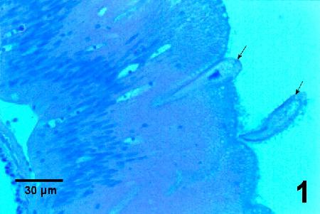
Figure 1. Type A ciliates (arrows) in the lumen of the gut and within a fold in the intestinal epithelium of Strongylocentrotus droebachiensis that were spawned and maintained in tanks for about 1½ years, Strait of Georgia, British Columbia, Canada. Haematoxylin and eosin stain.
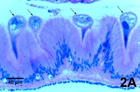
Figure 2A. Type B ciliates (arrows) within folds in the intestinal epithelium of Strongylocentrotus franciscanus that were spawned and maintained in tanks for about 1½ years, Strait of Georgia, British Columbia, Canada. Haematoxylin and eosin stain.
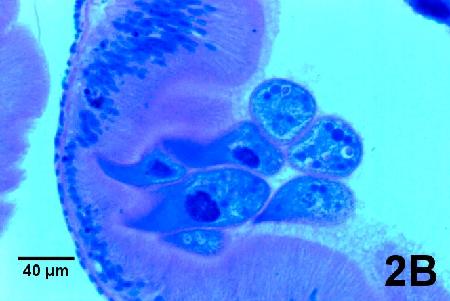
Figure 2B. a cluster of Type B ciliates in a fold of the intestinal epithelium of the same Strongylocentrotus franciscanus featured in Fig. 2A. Note the basophilic spheres in the cytoplasm of the ciliates furthest from the fold in the epithelium. Haematoxylin and eosin stain.
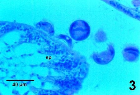
Figure 3. Trichodina sp. adjacent to the spine (sp) of Strongylocentrotus droebachiensis that were spawned and maintained in tanks for about 1½ years, Strait of Georgia, British Columbia, Canada. Haematoxylin and eosin stain.
Methods of control
Prevention and control impractical. These ciliates are ubiquitous.
References
Jangoux, M. 1990. Diseases of Echinodermata. In: Kinne, O. (eds), Diseases of Marine Animals. Volume III: Introduction, Cephalopoda, Annelida, Crustacea, Chaetognatha, Echinodermata, Urochordata. Biologische Anstalt Helgoland, Hamburg, Germany, pp. 439-567 (specifically see pgs. 454-457).
MacCallum, G.S., J. Blackbourn, S.E. McGladdery, S.M. Bower and J.T. Davidson. 2001. Disease issues relevant to the culture of shellfish in Atlantic and Pacific Canada. Bulletin of the Aquaculture Association of Canada 101-3: 5-12.
Citation Information
Bower, S.M. (2004): Synopsis of Infectious Diseases and Parasites of Commercially Exploited Shellfish: Ciliates Associated with Sea Urchins.
Date last revised: June 2004
Comments to Susan Bower
- Date modified: