
Kidney Coccidia of Mussels
On this page
Category
Category 2 (In Canada and of Regional Concern)
Common, generally accepted names of the organism or disease agent
Mussel kidney coccidia.
Scientific name or taxonomic affiliation
- Pseudoklossia semiluna in the family Aggregatidae Labbé.
- Pseudoklossia (=Hyaklossia) pelseneeri of the family Aggregatidae Labbé.
- Unidentified species of coccidia (Pseudoklossia-like).
Other species of coccidia have been described from the kidneys of oysters, scallops, clams and abalone from various locations around the world.
Geographic distribution
- Coast of British Columbia, Canada.
- East coast of the United States; and Galacia, Spain.
Host species
- Mytilus edulis/galloprovincialis/trossulus species complex.
- Mytilus edulis and Mytilus galloprovincialis.
Impact on the host
Infected kidney epithelial cells become hypertrophied and kidney tubules fill with coccidia. Heavy infections may cause kidney damage but associated mortalities appear restricted to artificial growing conditions. In British Columbia, among wild stocks of mussels, prevalence of infection was usually less than 16%, intensity of infection was usually light (less than 50 coccidia per histological section of kidney tissue), and evidence of associated pathology (apart from infected kidney epithelial cell) was not observed
Diagnostic techniques
Squash Preparations
Preliminary diagnosis can be made on the presence of large mature macrogamonts in squashes of kidney tissues.
Histology
Various stages of the coccidia can be observed in the cytoplasm of kidney epithelial cells. For P. semiluna, only gamontogonic and sporogonic and not merogonic developmental were observed, a characteristic of members of the family Aggregatidae. Mature macrogametocytes were crescent-shaped and oocysts sporulated within the host. Mature oocysts were spherical (23.9 ± 1.5 µm in diameter) with approximately 24 ellipsoidal sporocysts (about 6 by 3 µm), each of which contained 2 sporozoites.
Figures 1 to 3. Histological sections (haematoxylin and eosin stain) through the renal tissue of Mytilus sp. infected with various stages of Pseudoklossia semiluna.
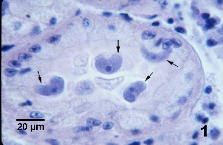
Figure 1. Four macrogamonts (arrows) within the cytoplasm of hypertrophied kidney epithelial cells. Note the crescent (moon) shape of this life stage from which the species name was derived.
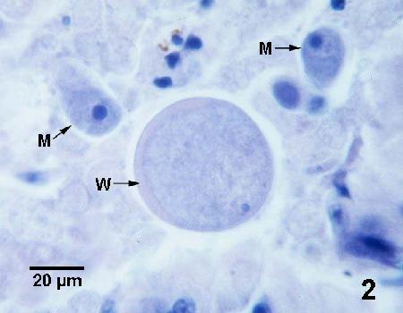
Figure 2. Unsporulated oocysts. Note the thickened portion (W) of the oocyst wall. Two developing macrogamonts (M) occur adjacent to this specimen.
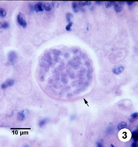
Figure 3. Sporulated oocyst with closely spaced sporocysts, each with two sporozoites. The thickened portion (arrow) of the oocyst wall is evident.
Methods of control
No known methods of prevention or control. However, species in the family Aggregatidae are heteroxenous and thus require another host (although none have been described to date) to complete the life cycle. Once the entire life cycle of these parasites are understood, management techniques may be feasible to eliminate the other host(s) and thus the parasite from the culture facility.
References
Bower, S.M. 1992. Diseases and parasites in mussels. In: E. Gosling (ed.) The Mussel Mytilus: Ecology, Physiology, Genetics and Culture. Developments in Aquaculture and Fisheries Science, Volume 25. Elsevier Press, Amsterdam, p. 543-563.
Bower, S.M. and A.J. Figueras. 1989. Infectious diseases of mussels, especially pertaining to mussel transplantation. World Aquaculture 20: 89-93.
Desser, S.S., S.M. Bower and H. Hong. 1998. Pseudoklossia semiluna n. sp. (Apicomplexa: Aggregatidae): a coccidian parasite of the kidney of blue missels, species of Mytilus, from British Columbia, Canada. Parasite 5: 17-22.
Farley, C.A. 1988. A computerized coding system for organs, tissues, lesions, and parasites of bivalve mollusks and its application in pollution monitoring with Mytilus edulis. Marine Environmental Research 24: 243-249.
Robledo, J.A.F., M.M. Santarém and A. Figueras. 1994. Parasite loads of rafted blue mussels (Mytilus galloprovincialis) in Spain with special reference to the copepod, Mytilicola intestinalis. Aquaculture 127: 287-302.
Villalba, A., S.G. Mourelle, M.J. Carballal and C. López. 1997. Symbionts and diseases of farmed mussels Mytilus galloprovincialis throughout the culture process in the Rías of Galicia (NW Spain). Diseases of Aquatic Organisms 31: 127-139.
Citation Information
Bower, S.M. (2001): Synopsis of Infectious Diseases and Parasites of Commercially Exploited Shellfish: Kidney Coccidia of Mussels.
Date last revised: June 2001
Comments to Susan Bower
- Date modified: