
Rickettsia-like and Chlamydia-like Organisms of Mussels
On this page
Category
Category 4 (Negligible Regulatory Significance in Canada)
Common, generally accepted names of the organism or disease agent
Rickettsia-like and Chlamydia-like organisms, Prokaryotic inclusion bodies (PIB).
Scientific name or taxonomic affiliation
Intracellular procaryote organisms belonging to the Rickettsiales.
Geographic distribution
Ubiquitous.
Host species
Mytilus trossulus, Mytilus galloprovincialis, Mytilus californianus, Mytilus edulis, and a wide variety of other marine bivalves including oysters, clams, cockles and scallops.
Impact on the host
Microcolonies in the epithelial cells of the gills and digestive gland. Infections are usually of light intensity, not associated with disease, and usually with no haemocytic response. However, large colonies can cause host cell hypertrophy with displacement and compression of the host cell nucleus against the basal membrane. An extreme example was provided by Gulka and Chang (1984) who reported large (up to 260 µm in diameter) basophilic inclusions of rickettsia-like organisms in the gill epithelium of M. edulis from Rhode Island, USA. The inclusions were surrounded by a host response consisting of an acidophilic layer (0.5 to 2.0 µm thick) of dense granular material containing round and spindle-shaped nuclei with granular amoebocytes located on the outermost periphery.
Diagnostic techniques
Histology
Basophilic microcolonies in cytoplasm of epithelial cells which may or may not be hypertrophied.
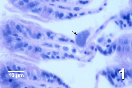
Figure 1. Large microcolony of Rickettsia-like organisms (arrow) in a hypertrophied epithelial cell near the distal tip of a gill filament of Mytilus trossulus from British Columbia, Canada. Haematoxylin and eosin stain.
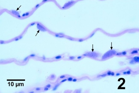
Figure 2. Several microcolonies of Rickettsia-like organisms (arrows) in the epithelial cells along the interlamellar junctions of gill filaments of Mytilus trossulus from British Columbia, Canada. Haematoxylin and eosin stain.
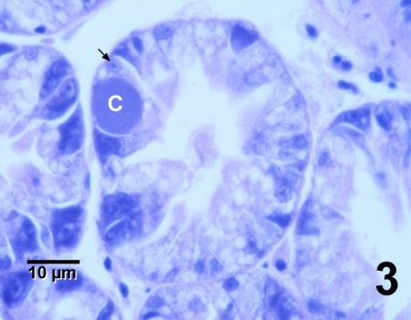
Figure 3. Microcolony of Rickettsia-like organisms (C), resembling a basophilic inclusion, in the cytoplasm of a normal-looking secretory epithelial cell, including nucleus (arrow), in a digestive gland tubule of Mytilus trossulus from British Columbia, Canada. Haematoxylin and eosin stain.
Methods of control
No known methods of prevention or control.
References
Bower, S. M. and A. J. Figueras. 1989. Infectious diseases of mussels, especially pertaining to mussel transplantation. World Aquaculture Review 20(4): 89-93.
Comps, M. 1983. Infections rickettsiennes chez les mollusques bivalves des côtes francaise. Rapports et Procès-verbaux des Réunions du Conseil International pour l'Exploration de la Mer 182: 134-136. (In French).
Gulka, G. and P.W. Chang. 1984. Host response to rickettsial infection in blue mussel, Mytilus edulis L. Journal of Fish Diseases 8: 319-323.
Harshbarger, J.C., S.C. Chang and S.V. Otto. 1977. Chlamydiae (with phages), mycoplasmas, and rickettsia in Chesapeake Bay bivalves. Science 196: 666-668.
McGladdery, S.E., R.E. Drinnan and M.F. Stephenson. 1993. A Manual of the parasites, pests and diseases of Canadian Atlantic bivalves. Canadian Technical Report of Fisheries and Aquatic Sciences No. 1931, p. 39-44.
Robledo, J.A.F., M.M. Santarém and A. Figueras. 1994. Parasite loads of rafted blue mussels (Mytilus galloprovincialis) in Spain with special reference to the copepod, Mytilicola intestinalis. Aquaculture 127: 287-302.
Villalba, A., S.G. Mourelle, M.J. Carballal and C. López. 1997. Symbionts and diseases of farmed mussels Mytilus galloprovincialis throughout the culture process in the Rías of Galicia (NW Spain). Diseases of Aquatic Organisms 31: 127-139.
Citation Information
Bower, S.M. (2004): Synopsis of Infectious Diseases and Parasites of Commercially Exploited Shellfish: Rickettsia-like and Chlamydia-like Organisms of Mussels.
Date last revised: December 2004
Comments to Susan Bower
- Date modified: