
Geoduck clam (Panopea generosa): Anatomy, Histology, Development, Pathology, Parasites and Symbionts
Developmental Stages of the Geoduck Clam
The development and morphology of the gametes of the geoduck clam are described on the web page pertaining to the Normal Histology of the Reproductive System.
Early developmental stages of the geoduck clam (days 0, 1, 2, 5, 7, and 14 after fertilisation of the ovum) were obtained from a commercial shellfish hatchery (Island Scallops Ltd., Qualicum Beach, B.C., Canada). These were relaxed (anaesthetized) for one hour in equal volumes of 15% solution of MgCl2 and sea water, then preserved for histology in Davidson’s solution. Specimens were imbedded in methacrylate plastic following the instructions provided by the supplier (JB-4® Embedding Medium, Polysciences, Inc. Warrington, PA 18976, USA). Sections of approximately 2 µm in thickness were cut with glass knives, then stained with Lee’s Methylene blue-basic fuchsin.
Larval size and morphological stage of development are dependent upon culture conditions. In the spring of 1996, about 21 days were required for geoduck clam larvae to reach metamorphosis and settle to the substrate.
Development of the Zygote
On day 0 the zygote underwent first and second cleavage divisions (Figs. 1 - 4). Gastrulation occurred on day 1 (Fig. 5) and on day 2, veliger larvae of about 116 µm had developed.
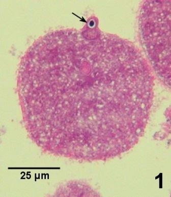
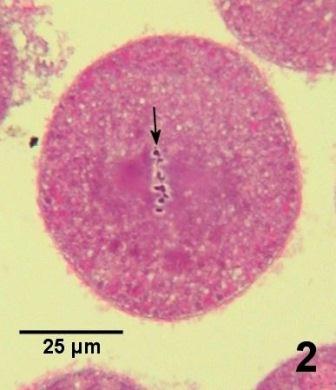
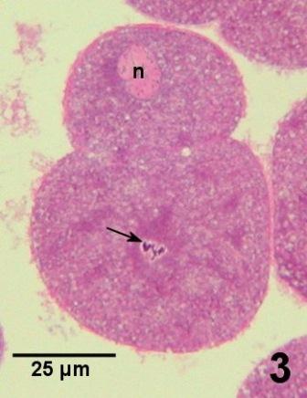
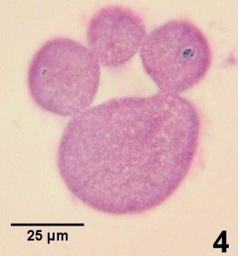
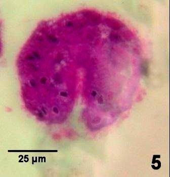
Figures 1 to 5. Development of geoduck clam zygotes within the first 36 hours after fertilization. Lee’s Methylene blue-basic fuchsin stain.
Veliger Larval Development
By day 2, about 60 hours after fertilization, the zygote had developed into veliger larvae about 116 µm in shell length. The anatomical terminology used in describing the location of structures of the veliger larvae is illustrated in Fig. 6.
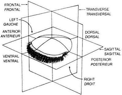
Figure 6. Orientation terminology for bivalve veliger larvae copied from Bower and Meyer (1990).
The characteristic feature of veliger larvae is the velum, a large ciliated organ that predominates the ventral surface of the larvae and is used for locomotion and the acquisition of food. The velum and associated cilia are retracted in all the veliger larvae illustrated histologically in this report. By day 2, the larvae had a shell length of about 116 µm and a well developed velum. Although the stomach and esophagus were evident, the other internal organs had not yet differentiated (Fig. 7a and b).
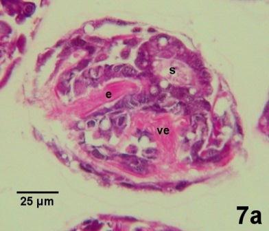
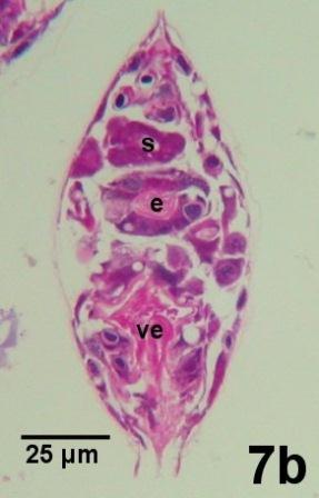
Figures 7a and 7b. Geoduck clam larvae on day 2 and at about 116 µm in shell length. Lee's Methylene blue-basic fuchsin stain.
By day 7 of development, the formation of the intestinal tract of the larvae was complete and the larvae had grown to a shell length of between 150 and 180 µm (Fig. 8a and 8b).
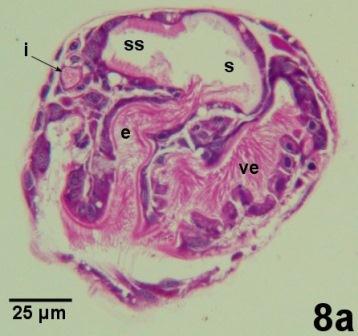
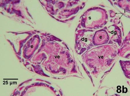
Figures 8a and 8b. Geoduck clam larvae on day 7 and at about 165 µm in shell length. Lee’s Methylene blue-basic fuchsin stain.
By day 14, the larvae had grown to between 226 and 244 µm in shell length. The apical organ, which may have a sensory function, has developed and is located dorsal to the velum. In addition, initial differentiation of some primordial cells suggests the early developmental stages of the gills and foot (Figs. 9a, 9b and 9c).
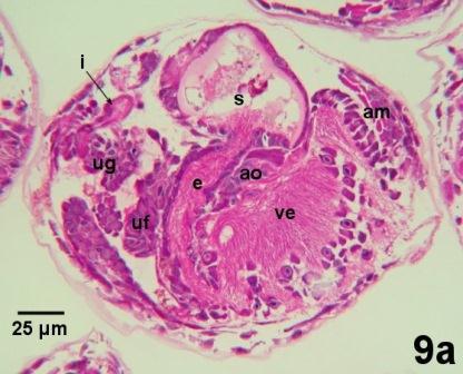
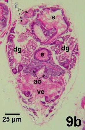
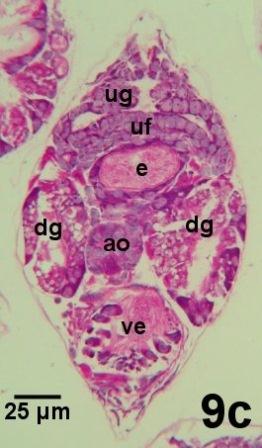
Figures 9a to 9c. Geoduck clam larvae on day 14 and at about 235 µm in shell length. Lee’s Methylene blue-basic fuchsin stain.
By day 17, the larvae were from 284 to 333 µm in shell length with evidence of the developing foot and associated byssal glands (Fig. 10). From this stage on, the swimming larva is usually referred to as a pediveliger. The apical organ was beginning to differentiate and eventually will form the cerebral and pedal ganglia with associated statocysts which will be used in orientation as the larvae metamorphoses from a pelagic to a sedentary life.
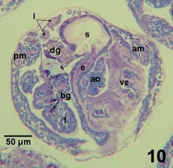
Figure 10. Sagittal section through a geoduck clam larvae on day 17 and at about 305 µm in shell length. The foot (f) with developing byssal glands (bg) is evident and the retracted velum (ve) takes less space within the larva in contrast to younger larvae. As in younger larvae, the digestive system (esophagus (e), stomach (s), digestive gland (dg) and intestine (i)) predominates the internal organs. However, the anterior (am) and posterior (pm) adductor muscles have developed and the apical organ (ao) is beginning to differentiate. Lee’s Methylene blue-basic fuchsin stain.
As the foot and gills of the pediveliger develop, the velum retracts (is absorbed). The larva can no longer maintain itself in the water column and settles to the bottom to find suitable habitat in the substrate. The early settlement stages, often referred to as spat, will grow into juveniles as they become established in the substrate.
References
Anderson, A.V. Jr. 1971. Spawning, growth, and spatial distribution of the geoduck clam, Panope generosa Gould, in Hood Canal, Washington. Ph.D. thesis submitted to University of Washington.
Barnes, Robert D. 1968. Invertebrate Zoology, 2nd ed. W. B. Saunders Company, Toronto, 743 pp.
Bower, S.M. and G.R. Meyer. 1990. Atlas of anatomy and histology of larvae and early juvenile stages of the Japanese scallop (Patinopecten yessoensis). Canadian Special Publication of Fisheries and Aquatic Sciences 111: 51 p.
Citation Information
Bower, S.M. and Blackbourn, J. (2003): Geoduck clam (Panopea generosa): Anatomy, Histology, Development, Pathology, Parasites and Symbionts: Normal Histology - Developmental Stages of the Geoduck Clam.
Date last revised: August 2020
Comments to Susan Bower
- Date modified: